Answer: a


Osteochondroma is the most common benign rib tumor and has a 3:1 male incidence. The stippled calcification and intact rib cortex are characteristic for this lesion in contrast to the bone destruction of Ewing sarcoma and combined bone destruction and “sunburst” calcification of osteogenic sarcoma. For both Ewing and osteogenic sarcoma, multimodality therapy using preoperative chemotherapy followed by resection yields better results than with radiation therapy. Osteochondromas in prepubertal children can be observed unless they become painful or enlarged, but are routinely resected in adults.
Answer: a, b, c


The posterior location of the infiltrate and fluid collection is typical of a parapneumonic empyema. The most important test is pleural aspiration which will usually yield frank pus, at which time a chest tube should be placed. Formerly, oily Dionosil was used to perform an empyemagram; this substance is now no longer commercially available. In the case of parapneumonic empyemas, tube drainage alone may be sufficient to allow full expansion of the lung. If this is not the case, a formal rib resection or early decortication should be performed. Decortication or marsupialization is indicated if the lungs fail to expand after 6–8 weeks. Every patient with spontaneous empyema should undergo bronchoscopy to rule out endobronchial obstruction by foreign body or tumor.
Answer: a, d, e
Skeletal chest wall defects that are full-thickness and occur posteriorly where they can be covered by the scapula do not require reconstruction. Anterior chest wall defects do require reconstruction, primarily to stabilize the chest wall and prevent paradoxical motion. The reconstruction should be immediate for optimal physiological benefit. Since Marlex mesh is porous, only a wound catheter is needed as pleural fluid will drain through it. PTFE, however, is a solid sheet necessitating both pleural and wound drainage.
Answer: a, e

Although the CT scan is a very sensitive indicator of pleural effusion

, a lateral decubitus is the simplest way to differentiate fluid from pleural thickening or fibrosis. Tuberculous pleuritis is difficult to diagnose by stain or culture which have a 30% yield, but the diagnosis is facilitated by needle biopsy of the pleura. Pleural fluid glucose lower than in serum is characteristic of rheumatoid arthritis, neoplasms, and tuberculosis as well as empyema. A red-tinged fluid can occur from needle trauma, but even frankly bloody fluid in this patient may reflect trauma as well as underlying malignancy. Pleural inflammation induces reactive changes in mesothelial cells that makes them resemble lymphocytes, so a lymphoma diagnosis is suspect.
Answer: e


Although mediastinal granulomatous disease is one cause of the superior vena cava syndrome described, the most common cause (75%) is malignant disease. A venogram adds little information to the typical findings and increases risk from extravasation of contrast medium subcutaneously from the venous hypertension. Mediastinoscopy can be used for diagnosis with recognition of increased risk of bleeding and airway problems from the edema associated with the endotracheal intubation required for the procedure. If a malignancy is found, operative resection is usually precluded by the extent of mediastinal invasion. Fortunately, in the case of benign disease, the symptoms tend to improve with time as chest wall and mediastinal collaterals enlarge.
Answer: d, e

The pectoralis major

muscle can be used for reconstruction but the medial and lateral pectoral nerves are named from their respective cords of the brachial plexus. The serratus anterior muscle holds the scapula to the chest wall and its absence produces the functional and cosmetically disabling winged scapula. The serratus posterior muscle is attached to the 7th cervical and first three thoracic vertebrae posteriorly and functions as an accessory muscle of respiration. The constancy of the vascular pedicle to the latissimus dorsi and its size allow this muscle to be used to reconstruct defects of the head, neck, chest wall and pleural cavity. It is innervated by the thoracodorsal nerve with fibers from C6, C7 and C8.
Answer: d, e


The posterior mediastinal location of the tumor is most indicative of a neurogenic tumor while teratomas are characteristically found in the anterior mediastinum. Neurogenic tumors can undergo malignant degeneration and should be resected, particularly in this symptomatic patient even if known to be present for years. The radicular pain suggests the possibility of intraspinous extension of the tumor, and therefore a neurosurgical consultation is appropriate. Both urinary vanillylmandelic acid elevation and vasoactive intestinal polypeptide can be produced by ganglioneuroma but would not be characteristic of a paraganglioma.
Answer: ABCD
DISCUSSION:


The single-breath DLCO is a screening test that has been shown to be decreased in all of the above examples. It is an estimate of the total capacity of the functional alveolar microarchitecture and has been demonstrated to be an independent measure of physiologic capability apart from the FEV 1 and forced ventilatory capacity.
Answer: ABCDE
DISCUSSION:


The most important clues to impairment of respiratory function are found in the history and physical examination. A negative history and physical examination in combination with a relatively normal room air arterial blood gas and normal chest film are sufficient to screen patients to support the clinical impression that there is minimal pulmonary disease. Patients with symptoms, positive physical findings, and/or abnormalities in the arterial blood gases or chest film can be screened most effectively with an additional evaluation of the vital capacity and FEV 1. More elaborate tests such as cardiopulmonary exercise testing are reserved for patients with obvious and marked impairment of pulmonary function who are being evaluated for the feasibility of surgical intervention.
The Correct Answer: C
DISCUSSION:
Pulmonary artery circulation transports oxygenated blood to the alveoli level where gas exchange occurs, and it is here that the matching of ventilation and perfusion is so important during the postoperative period. The loss of lung volume that generally occurs after all surgical procedures does not return to baseline for 5 to 7 days and may play an important role in the ventilation-perfusion ratio. Improving or returning lung volume to normal is performed by manipulating functional residual capacity (FRC) and preventing atelectasis, which in turn maintains circulation to the alveolus and optimizes the ventilation-perfusion ratio.
The Correct Answer: A
DISCUSSION:


Chronic bronchitis may have an acute component, and in these patients therapy with antibiotics and bronchodilators may improve the flow rate as measured by pulmonary function tests within 3 or 4 days of the cessation of smoking and treatment of the acute condition. However, the chronic bronchitic will continue to produce large amounts of mucus, most evident in the morning, even after the acute process has been resolved. Clearance of these secretions is hampered by the inability to cough, perhaps secondary to the pain of thoracotomy or abdominal surgery or by a decrease in the number of ciliary cells that help move mucus up the tracheobronchial tree. This causes plugging of small airways and atelectasis, which may progress to pneumonia. For this reason, cessation of smoking for 3 to 5 days before surgery is very beneficial in preventing pulmonary complications during the postoperative period.
The Correct Answer: DDISCUSSION: The bronchial circulation is the primary blood supply for the conducting airways, pulmonary vessels, lymphoid tissue, and squamous cell carcinomas. In conditions such as mitral stenosis, bronchiectasis, or chronic obstructive pulmonary disease, the rich peribronchial venous network that drains the bronchial circulation may expand considerably, creating significant left-to-right shunts. Whenever the pulmonary artery circulation is obstructed, there is a tendency for bronchial circulation to increase; thus, the bronchial circulation is an important consideration during lung transplantation as well as in the surgical treatment of cyanotic congenital heart disease and chronic pulmonary embolism.
The Correct option is A
Cor pulmonale
This disorder is defined as a disturbance of structure and function of the right ventricle as a result of precapillary pulmonary hypertension in chronic lung disease.
The most common cause is chronic obstructive lung disease (COLD ).
Right ventricular failure may resulting in prominent edema in the lower extremities and perhaps ascites.
More often we find normal ejection fraction of the RV at rest and even during exercise and normal glomerular filtration fraction. In these patients edema is a result of hypercapnea, which leads to increased reabsoption of bicarbonates followed by pasive reabsorption of sodium chloride and water in the kidney.
Also chronic hypoxemia and consequent vasoconstriction in renal vascular bed and reduction of sodium excretion play a role.
The Correct option is A
The use of thrombolytic drugs also raises the potential for intramyocardial hemorrhage, which would increase the volume and pressure on the poorly healing heart; this overload can cause the heart to rupture. One particular study on the relation between thrombolysis and rupture is the Late Assessment of Thrombolytic Efficacy (LATE) study by Becker and associates. The study demonstrated that the group receiving rt-PA within 6 to 12 hours of symptom onset had the highest number of ruptures, but the data were not statistically significant.
The Correct option is
COn July 21, 1768, William Heberden presented
"Some Account of a Disorder of the Breast" to the Royal College of Physicians, London:
"But there is a disorder of the breast marked with strong and peculiar symptoms, considerable for the kind of danger belonging to it, and not extremely rare. The seat of it, and sense of strangling and anxiety with which it is attended, may make it not improperly be called angina pectoris."
Heberden appropriated the term angina from the Latin word for strangling. His classic account marks the beginning of our appreciation of coronary artery disease.
The correct option is A
Serous Tumors —"serous" from yellow fluid in cysts; elevated serum CA 125
The Correct option is
CYolk sac tumor (endodermal sinus)
- - solid mass w/ hemorrhage and necrosis w/ Schiller-Duval Bodies.
- Hyaline droplets contain AFP
- Produce alpha fetoprotein
- Rapid growing and aggressive – surgery and chemo
The Correct option is B
Choriocarcinoma – produce hCG and are widely metastatic
The Correct option is
DGranulosa Cell tumors:
- These secrete estrogen, so they may cause abnormal vaginal bleeding or endometrial hyperplasia/carcinoma.
- The tumor itself has malignant potential too.
- Pathology includes “Call-Exner bodies” (small red cavities) and “coffee bean” cells.
- Occurs any time after puberty.
The Correct option is A
Neisseria gonorrhoeae is recognized as a pathogen in any human clinical specimen in which it occurs. Its presence is indicative of a reportable sexually transmitted disease. Genital, oropharyngeal or anorectal specimens inoculated onto selective enriched medium (modified Thayer-Martin) and incubated at 35-37oC in 5% CO2 overnight are suitable specimen.
The Correct option is
CThis patient has a
Complete hydatidiform mole.
The placenta will show:
Gross: - Distended, edematous villi ("bunch of grapes")
- No gestational sac (usually)
- Amount of tissue variable – classically large
Micro: - Uniformly edematous (hydropic) villi with dissolution of central stroma (cavitation/cistern)
- Villous vessels absent (usually)
- Trophoblastic hyperplasia – circumferential, haphazard, involves CT/ST/IT
- Trophoblastic atypia
The Correct option is
D
Toxemia of pregnancy is a clinical syndrome characterized by elevated blood pressure, protein in the urine, fluid retention and increased reflexes. It occurs only during pregnancy and resolves completely after pregnancy. It is seen most often as women approach full term, but it can occur as early as the 22nd week of pregnancy. It's cause is unknown, but it occurs more often in:
- Women carrying their first child
- Multiple pregnancies
- Pregnancies with excessive amniotic fluid (polyhydramnios)
- Younger (<17) and older (>35) women
The Correct option is C
Human Parvovirus is the etiologic agent causing erythema infectiosum (Fifth Disease), a common pediatric illness. Maternal infection has been associated with hydrops fetalis and fetal death.
Anemia in newborn infants is a common manifestation of hemolytic disease or congenital parvovirus B19 infection. Parvovirus infection of erythroid precursors in the bone marrow leads to sudden cessation of erythropoiesis. Duration of Parovirus infection is 7 – 10 days, but mature red cells live for about 10 days, so hemoglobin falls suddenly. The RBC’s are normal size, but the erythroid precursor is the “size of a Volkswagen, absolutely enormous,” which is typical of parvovirus infection.
The Correct option is E
Hamartoma's are abnormally developed native tissue. They are always benign.
The Correct Option is A
Various congenital anomalies are known to be associated with Wilms' tumor. Aniridia, hemihypertrophy, genitourinary tract anomalies (eg, cryptorchidism and hypospadias), Beckwith-Wiedemann syndrome, and Denys-Drash syndrome have been reported to confer an increased risk of the development of Wilms' tumor
The Correct option is B
An important characteristic of teratogens is that they work during specific critical periods.
During the pregastrulation period of development (cleavage, blastocyst, early primitive streak) the embryo is refractory to teratogenesis but very sensitive to toxins.
As gastrulation and organogenesis begin, the embryo becomes selectively susceptible to most teratogenic agents.
The critical period is that time when a given organ system or tissue exhibits its greatest sensitivity to a particular teratogen. This period usually corresponds to the time of its most dramatic growth and morphogenesis.
The heart, for example, is most sensitive during the third to fifth weeks, while the critical period for the external genitalia is during weeks eight and nine.
The brain appears to be sensitive to teratogens from the beginning of the third week until the end of pregnancy.
The Correct Option is E
Bronchoalveolar lavage is similar to a transtracheal aspirate in that a small volume of sterile fluid is instilled into the airways and then re-aspirated in hopes of retrieving material from airway secretions and lesions. The difference is that with bronchoalveolar lavage, the lower parts of the airways are sampled. Bronchoalveolar lavage (BAL) is particularly helpful in patients in whom the cause of pulmonary disease or infection is not known. Depending upon the nature of the material obtained by bronchoalveolar lavage, we may need to culture some of the material in order to definitively determine whether or not there are bacteria or fungi present.
The Correct Answer is
ASerous cystadenocarcinomas account for 40% all ovarian cancers
- most common malignant ovarian tumor
- Papillary serous carcinomas are managed as grade 3 carcinomas irrespective of their histologic profile
The Correct option is
BPattern of Spread of Cervical Carcinoma:- Begins on Cervix – often identifiable by physical exam/pap smear
- Grows locally
- Can grow into the lower uterine segment, obstructing the uterus
- Extends to pelvic sidewalls, can involve distal ureters as they course into Bladder
- Can involve bladder, rectum
- Pelvic lymph node chains, more extensive disease extends to para-aortic node
- Distant metastases, liver, lung
The Correct option is
BPaget's disease of the vulva usually presents as a scaly, erythematous lesion on the vulva and in most cases represents a metastatic, intra-epidermal adenocarcinoma arising from an underlying sweat gland carcinoma.
Micro:- Neoplastic elements are glandular cells
- Large, pale cells; single or nested - occasional glands.
- Abundant cytoplasm - vacuoles may be present.
- Concentrated in suprabasal portion of epidermis.
- Pilosebaceous apparatus frequently involved.
- May be multifocal with intervening normal epidermis
The Correct option is D
The histopathologic types of cervical carcinomas show that squamous cell carcinoma accounts for 80% cervical carcinomas. Adenocarcinomas for 15%. And 5% other types.
Adenocarcinoma has the same risk factors, but appears to have a stronger association with oral contraceptives than does the squamous cell cancer.
The strong association between HPV infection and cervical neoplasia has been established for invasive carcinoma, its precursor lesions and adenocarcinoma in situ. Therefore, the major factors associated with the development of cervical cancer are the HPV infection and its associated subtypes (high-risk vs. low-risk) and their persistence.
The Correct option is C
A cone biopsy is aimed to diagnosis of the lesions. Nevertheless, in most cases this procedure will also have a therapeutic value (whenever the whole lesion is included in the specimen and the surgical margins are lesion-free). Hysterectomy will be necessary if the surgical margins of a cone biopsy are affected by the lesion.
The Correct option is C
Decidual cells - Cells of the endometrial stroma within the wall of the uterus differentiate into decidual cells in response to the implanting embryo by accumulating lipid and glycogen.
Decidual reaction - In response to implantation, endometrial stromal cells accumulate lipid and glycogen, the endometrium thickens, the endometrial glands enlarge, and endometrial veins and spiral arteries make connections with trophoblastic lacunae.
The Correct option is
D- Endometrial polyps are localized hyperplastic overgrowths of endometrial glands and stroma that form a sessile or pedunculated projection from the surface of the endometrium.
- They develop as solitary or multiple soft tumors.
- Diffuse endometrial hyperplasia may consist of multiple polypoid projections.
- In most cases the polyp is made up of endometrium similar to that seen in the basalis and does not show secretory changes.
- Less than one-third of the polyps contain functional endometrium, similar histologically to the endometrium from which they arise.
- Polyps frequently show a microscopic picture of cystic hyperplasia; less commonly they show one of adenomatous hyperplasia.
- The tip of the polyp may be necrotic and inflamed, particularly if it is long and protrudes into the cervix.
- Squamous metaplasia of the lining surface has been observed.
The Correct option is
B- Fibrocystic breast changes are the most common cause of breast tenderness and breast lumps.
- Fibrocystic disease is most prevalent in middle-aged women.Fibrocystic change does not occur prior to puberty, and it is unusual to diagnose its onset after menopause.
- Fibrocystic change of the breast refers to a constellation of morphological features characterized by
(1) cystic dilation of terminal ducts,
(2) relative increase in fibrous stroma, and
(3) variable proliferation of terminal duct epithelial elements
The Correct option is
BSeminoma- Malignant germ cell tumor
- Most common germ cell tumor (40%)
- Peak in mid 30’s
- See, painless enlarged testis
- Assoc. w/ increased hCG
- Very radiosensitive and can usually be cured
The Correct option is E
Discussion: Adenocarcinoma of prostate- Peak age - 65-70 80%
- arise in peripheral portion of gland;
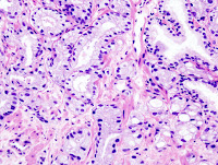
- small acini glands with single layer, prominent nucleoli, neoplastic acini invades stroma
- Gleason grading: 2-4 well differentiated, 5-7 moderate, 7-10 poor;
- Staging: A (microscopic), B (nodules confined to gland), C (local invasion beyond prostate), D (distant metastasis);
- prostate specific antigen (PSA) and acid phosphatase are markers
The Correct option is D
Vulvar lichen sclerosis et atrophicus results in marked hypopigmentation with tissue thinning and scarring.Lichen sclerosis et atrophicus may be asymptomatic until the patient has sufficient scarring to cause complications such as dyspareunia, dysuria, and difficulty voiding. With continued progression, the clitoris, the labia minora, or even the opening of the vagina may be obliterated. Simple inspection of the external genitalia can lead to early diagnosis and treatment to avoid such serious complications.
Chronic vulval dystrophies are not precancerous lesions, providing there is no dysplasia- there is no a higher risk for carcinoma
The Correct option is
DEndocervical Polyps
- Innocuous, occur in 2-5% of adult women, measure up to 5 cm.
- May cause spot bleeding. Micro = inflamed mucous secreting glands
- Excision is curative
The Correct option is C
It is the interaction of oncoproteins E6 and E7 with various cellular proteins that immortalizes HPV cells in high-risk HPV. Two of HPV’s oncoproteins, E6 and E7, interact with two tumor suppressor cellular proteins, p53 and pRb respectively. When E6 interacts with p53, p53 is degraded resulting in the immortalization of HPV cells because p53 can no longer monitor the cellular repair process.
E7 evolved from the same common ancestor as E6 and interacts with p105Rb which allows the release of E2F transcription factors and activates genes that promote the cell cycle. Additionally, E7 also blocks negative cell cycle regulators like cyclin-dependent kinase inhibitors. Furthermore, E7 is a mitotic mutator, meaning that it induces centrosome abnormalities to generate aneuploidy. Like E6, E7 is present continuously during carcinogenesis.
The Correct option is D
1st evidence of ovulation is the subnuclear vaculoation in the cytoplasm of the endometrium epithelium.
The Correct option is E
The endometrial stromal sarcoma may be pure or mixed (most of them are pure), and the stroma is invariably homologous.
The Correct option is B
Bloody discharge most often results from an intra-ductal papilloma, it may mark an underlying cancer
The correct option is
EChoriocarcinoma
- A malignant tumor derived from trophoblast. 50% come from complete hydatidiform moles.
- There are no villi at all! By definition, if there are villi it is hydatidiform mole, not choriocarcinoma.
- The tumor is a hemorrhagic nodule with central necrosis, because the choriocarcinoma has no intrinsic blood supply (as all other cancers do). So only the outer rim of the tumor is viable.
- The tumor recapitulates endovascular trophoblastic migration, with syncytiotrophoblasts around cores of cytotrophoblasts. Highly destructive, infiltrative growth.
- Serum hCG is mildly elevated, but hCG is extremely high inside tumor cells.
- Choriocarcinoma metastasizes hematogenously, yet has a very good prognosis with chemotherapy.
The Correct option is
B | | Pathologic Features | Pathogenesis | Complications/Fetal Outcome |
| Succenturiate lobe | Commonest shape variation in which one or more discrete lobes separated from main placental mass. Cord inserts into main placental mass. Vascular supply reaches accessory lobe by running through fetal membranes unsupported by underlying villous tissue. | ? | Vessel rupture during delivery fetal blood loss Retention of lobe postpartum bleeding, infection Vessel thrombosis - possibly associated with other fetal thrombi |
| Placenta "creta" | Placenta accreta, increta, percreta Abnormally adherent placenta - does not separate from uterus after delivery of fetus Variable degrees of abnormal adherence or invasion of myometrium common
| Pathogenesis - lack of decidua (decidua normally limits invasive properties of trophoblast) | Complications - failure of placental separation life threatening post partum bleeding - antepartum bleeding - uterine rupture |
The Correct option is B
Twin to Twin Transfusion Syndrome (TTTS) is a disease of the placenta. It affects identical twins during pregnancy when blood passes disproportionately from one baby to the other through connecting blood vessels within their shared placenta. One baby, the recipient twin, gets too much blood overloading his or her cardiovascular system, and may die from heart failure. The other baby, the donor twin, does not get enough blood and may die from severe anemia. The babies are normal. The abnormalities are in the placenta.
The Correct Answer is E
In preeclampsia, the diseased placenta produces a circulating factor, which affects the maternal endothelium. Two gross abnormalities commonly seen in pre-eclamptic pregnancies result from these vascular abnormalities. The reduction in luminal diameter caused by the absence of physiological vascular changes, vasospasm, persistent intraluminal endovascular trophoblast, acute atherosis and thrombosis can lead to a markedly impaired blood flow through the spiral arteries. This results in placental infarction.
The Correct option is
EFetal complications of maternal diabetes
- Congenital anomalies
- Congenital heart disease (VSD, transposition of the great arteries)
- Neural tube defects
- Caudal regression
- Macrosomia, intrauterine growth restriction (IUGR)
- Stillbirth
The Correct option is B
Isotretinoin exposure is characterized by central nervous system defects (eg, hydrocephalus, microcephaly, structural errors of neuronal migration), but not meningomyelocele. Other associated abnormalities include facial asymmetry, microtia or anotia, conotruncal cardiac abnormalities, and mental deficiency. The risk for retinoic acid embryopathy is 35% in women who continue to ingest the agent past the 15th day following conception.
The Correct option is
B- Wilms' tumor is the most common childhood renal tumor, accounting for approximately 6% of all pediatric malignancies.
- Cytogenetic analysis of children who have the WAGR syndrome demonstrated deletions at chromosome 11p13
- In addition to the two genetic loci on chromosome 11, familial Wilms' tumor predisposition at FWT1 (17q) and FWT2 (19q) loci has been identified
The Correct option is
ANeuroblastoma is a form of cancer, found primarily in children, that attacks the developing nerve cells.
Neuroblastoma is the most common extracranial solid tumor in infants and children, accounting for 6% to 10% of all childhood cancers and 15% of all pediatric cancer deaths in the United States.
- Approximately 30% of primary neuroblastomas demonstrate N-myc amplification, which is strongly correlated with advanced-stage tumors and poor prognosis, independent of patient age and staging
- Nonmorphologic favorable prognostic indicators are age less than 1 year, clinical stages 1, 2, and 4S, and N-myc nonamplification.
- Neuroblastomas arise from primordial neural crest cells, which migrate during embryogenesis to form the adrenal medulla and sympathetic ganglia.
- As a result, neuroblastomas occur in the adrenal medulla or anywhere along the sympathetic ganglia, most notably in the retroperitoneum and posterior mediastinum.
- Plain radiographs and ultrasonography are routinely obtained for patients who have any suspicious mass. Chest radiographs revealing posterior mediastinal mass can narrow the differential diagnoses; calcification is also detected in up to 50% of cases.
Answer: C
DISCUSSION:
The treatment of every comatose patient begins with an assessment of the patient's respiratory system, followed shortly thereafter with an assessment of the patient's cardiovascular system. The unconscious patient's normal protective pharyngeal reflexes are compromised, making mechanical airway obstruction and aspiration pneumonia common events. Hypotension, secondary to intra- or extracorporal hemorrhage, is deleterious to the patient's cerebral injury. Neurologic assessment is undertaken only after the patient's respiratory and cardiovascular status are secured.
Answer: A,B
DISCUSSION:
An epidural hematoma is a blood clot situated between the skull and the dura. Epidural hematomas are usually arterial in origin and most often are secondary to hemorrhage from the middle meningeal artery. Approximately 90% of adult patients with an epidural hematoma have a concomitant skull fracture. Such skull fractures are much less common in children under the age of 2 years. The epidural hematoma is best diagnosed before transtentorial herniation and the development of third cranial nerve palsy (“blown pupil”). The outcome of therapy is directly related to the patient's level of consciousness before surgery. The clinical diagnosis of an epidural hematoma is rarely confirmed by brain CT.
Answer: D
DISCUSSION:
In the past, the preferred treatment of a brain abscess was total surgical excision. Now that such abscesses can be followed closely by CT, aspiration and drainage is usually employed, at least initially, to reduce the mass effect, provide information about the pathogens, and lower the risk of intraventricular rupture while the abscess is treated by systemic administration of antibiotics.
Answer: D,E
DISCUSSION:
Extradural neoplasms are usually malignant, the most common type being a metastasis to a vertebra from a primary carcinoma elsewhere in the body. A meningioma is an extramedullary tumor arising from the meninges surrounding the spinal cord rather from within the cord itself. Most intradural extramedullary neoplasms are benign tumors (meningiomas, neurofibromas, schwannomas) that are treated by surgical excision without postoperative radiotherapy. Despite its name, the hemangioblastoma is a benign tumor. It typically arises within the spinal cord and can be cured if it is completely removed surgically.
Answer: A,B,C,D,E
DISCUSSION:
Intraspinal dermoid and epidermoid tumors and lipomas are benign lesions that can be found within the subarachnoid space or the spinal cord, or both. They are most common in the lumbosacral area. Dermoid and epidermoid tumors can be associated with spinal dysraphism and in particular with a dermal sinus tract that opens onto the back, usually in the lumbosacral region. Lipomas are also associated with spinal dysraphism, at times in the form of a lipomyelomeningocele with a tethered spinal cord.
Answer: A,B,C
DISCUSSION:
Most symptomatic lumbar disc herniations do occur in a posterolateral direction, impinging on the overlying nerve root. About 95% of lumbar disc herniations occur at the L5–S1 or L4–L5 level. Approximately 4% occur at the L3–L4 level, and less than 1% at the L2–L3 or L1–L2 level. Sciatica is a term used to refer to pain along the course of the sciatic nerve. A ruptured lumbar disc typically causes low back pain and ipsilateral sciatica. The mechanical signs of a lumbar disc herniation include paravertebral muscle spasm, lumbar scoliosis, tenderness over one or more of the lower lumbar spines, limitation of low back motion, limitation of straight leg raising, and a positive popliteal compression test. Weakness of dorsiflexion of the foot is a neurologic sign, not a mechanical sign. Plain x-ray films of the spine do not demonstrate the presence and location of a lumbar disc herniation except in the rare instance of a calcified disc herniation. Myelography, CT, or MRI is needed to visualize the herniated disc.
Answer: A,C
DISCUSSION:
A lumbar disc herniation at the L5–S1 or L4–L5 level typically causes low back pain and ipsilateral sciatica. If a ruptured L5–S1 disc causes weakness, it ordinarily involves plantar flexion of the ipsilateral foot. Although a diminished or absent ankle jerk can be caused by either an L5–S1 or an L4–L5 disc herniation, it is more common with the former. The L5–S1 disc herniation ordinarily affects the S1 nerve root, which supplies the lateral aspect of the foot, including the small toe.
Answer: B,E
DISCUSSION:
A symptomatic cervical disc herniation usually occurs in a posterolateral direction, although a directly posterior (central) herniation may occasionally occur. The posterolateral herniated disc can be removed by either a posterior or an anterior approach, but the anterior approach is preferred for the central herniation because the surgeon can remove the ruptured disc without manipulating (and possibly injuring) an already compromised spinal cord. Cervical spondylosis represents a combination in the cervical spine of degenerative disc disease and osteophyte formation (including that from osteoarthritis of the apophyseal joints and the joints of Luschka).
The cervical spine contains the joints of Luschka, which are not present elsewhere in the spine. These joints, one on each side of the disc, are separate from the more posteriorly situated facet joints (apophyseal or interpedicular joints). The term cervical myelopathy refers to dysfunction of the cervical portion of the spinal cord. Pain and/or neurologic dysfunction in the distribution of one or more cervical nerve roots is termed cervical radiculopathy. Neck movement, especially extension, often intensifies the neck and arm pain of a patient with a cervical disc herniation.
Answer: A,C,E
DISCUSSION:
Fascicles within a peripheral nerve do divide and recombine along their course, forming funicular plexuses. If a segment of a nerve is removed and the remaining ends are reapproximated, the fascicles will not match exactly. In neurapraxia (first-degree nerve injury) anatomic continuity of the axons is preserved, but there is selective demyelination. Surgical repair is not necessary. Recovery does not depend on regeneration and occurs within days or weeks. With neurotmesis there is significant disorganization in the nerve or actual disruption of its continuity, which precludes recovery without surgical repair. Axonal sprouting ordinarily begins 10 to 20 days after transection of a peripheral nerve. The patient's age affects the rate and success of nerve regeneration: the younger the patient is, the faster and more complete is the recovery.
Answer: D
DISCUSSION:
This patient has all of the neurologic components of the most common cervical disc syndrome, that caused by a herniation at the C6–C7 level with compression of the C7 nerve root.
Answer: C
DISCUSSION:
The Hoffmann-Tinel sign identifies the most distal point of small nerve fiber regeneration. As nerve regeneration progresses, this point moves farther away from the level of the nerve injury. Causalgia is a specific severe pain syndrome that may accompany a partial injury to a mixed peripheral nerve. As compared with primary repair, the extent of damage to a nerve can be better assessed and the correct amount trimmed off, with a secondary repair 3 to 8 weeks after the injury; the epineurium and perineurium are stronger and can be sutured more easily; optimal operating room conditions can be arranged; and there is no time for wallerian degeneration (i.e., the involved neurons are capable immediately of regenerating new distal segments, and the regenerating axons can penetrate the repair site before a significant amount of scar forms). If a clinically non-functioning nerve is in continuity when it is explored some weeks after the initial injury the surgeon may find it helpful to stimulate the nerve electrically proximal to the injury and to look distally for evidence of muscle contraction or transmission of nerve action potentials. If there is no evidence of transmission across the area of injury, the injured portion of the nerve should be excised and the cut ends sutured together. If there is transmission across the area of injury, surgical treatment should be limited to external neurolysis. A disrupted nerve should be re-approximated surgically, but only after each end has been trimmed back to healthy fascicles. The trimmed nerve ends must not be under tension when they are sewn together.
Answer: E
DISCUSSION:
Café-au-lait spots are not a feature of
spina bifida occulta
. The other four skin features all may be associated with significant intradural pathology and warrant further investigation, most commonly with magnetic resonance imaging (MRI). A dermal sinus tract that overlies the coccyx is a pilonidal sinus and is not likely to be associated with intradural pathology.
Answer: E
DISCUSSION:
Myelomeningoceles are usually associated with hydrocephalus and the
Chiari II malformation
. The myelomeningocele sac is a midline dorsal spinal mass associated with defects in the skin, bone, and dura overlying the neural placode, and the sac is readily apparent at birth. Although the innervation of the bladder is dysmorphic, the majority of patients can achieve social urinary continence through the use of clean intermittent bladder catherization.
Answer: AC
DISCUSSION:
Horner's syndrome is due to loss of sympathetic innervation to the head and neck and includes ptosis, anhidrosis, miosis, and the appearance of enophthalmos. The pupil is small owing to loss of the tonic dilating effect of the sympathetics in the presence of continued parasympathetic activity. There is sympathetic innervation to Muller's muscle in the upper lid. Sympathetic nerves supply the sweat glands. It commonly follows stellate ganglion resection and involves removal of the T1 cord level sympathetic outflow.
Answer: AC
DISCUSSION:
Cordotomy results in a lesion of the spinothalamic tract, which is a crossed pathway carrying signals for pain and temperature.
Answer: AB
DISCUSSION:

Because seizure surgical procedures can never be guaranteed to alleviate seizures, it is only undertaken when

medical therapy fails to control the patient's seizures at doses that do not produce intolerable side effects. Most surgical procedures are aimed at removing a single epileptogenic area of cerebral cortex and are rarely employed in patients with multiple areas of epileptogenic cortex. Eloquent areas of cerebral cortex such as those subserving speech or hand functions generally are not intentionally resected in an attempt to achieve seizure control.
Answer: B,C,D
DISCUSSION:
Since the exact anatomy of an epileptogenic focus remains obscure, the focus of the patient's seizures is determined by concordance of the clinical manifestations of the seizures, abnormalities demonstrated by cerebral imaging, and abnormalities demonstrated by electroencephalography.
Answer: C
DISCUSSION:
The biopsy of a lesion, radiotherapy treatment of an arteriovenous malformation, and excision of an arteriovenous malformation are all procedures for structural lesions of the brain that can be imaged by either CT or MRI. These structural lesions may or may not cause neurologic changes, but the treatment directed at them is intended principally to keep lesion-induced damage from increasing (for example, with the development of hemorrhage). On the other hand, the thalamus is expected to have a normal structural appearance and function in Parkinson's disease, when the neurochemical abnormality is located in the substantia nigra and the striatum (caudate and putamen). Thus, a lesion is made in the thalamus principally to affect the function of the brain, altering a normal component of one of the motor circuits to compensate for the changes in the other parts (i.e., the basal ganglia).
Answer: B
DISCUSSION:
Frame-based and frameless procedures both use digitized imaging studies as the basis for converting the scan coordinate system into a treatment coordinate system. Both types of procedures also require rigid fixation of the patient's head to the operating room table and can be performed in the presence or absence of a lesion. The critical difference is the use of a rendered, three-dimensional image and the three-dimensional digitizer, which together allow the alignment to be generated between the patient's imaging studies and the patient; this alignment occurs in frame-based stereotactic procedures because of the imaging study performed after the frame is applied.
Answer: b, d, e
A brain abscess is a purulent lesion of brain tissue, beginning as a focal infection, usually in the white matter surrounded by a typical inflammatory response. Brain abscesses usually are secondary to focal infection elsewhere. Abscesses that develop by direct intracranial extension are usually solitary and are typically found in the frontal and temporal lobes. Multiple brain abscesses that develop in the septic patient are often related to bacterial endocarditis, pneumonia, and diverticulitis. Abscess formation is frequent among patients with compromised immunity either from an underlying illness or during pharmacologic immunosuppression (i.e., during organ transplantation). Signs and symptoms of brain abscess are related to its mass effect. Headache, focal neurologic deficits, and impaired mentation are often noted. There may be little or no evidence of infection and the patient may be afebrile. Seizures may occur. Intracranial epidural abscesses are quite uncommon and are usually caused by a local extension of osteomyelitis or by hematogenous spread from a distant suppurative focus.
In cases of early abscess formation or high surgical risk, medical therapy alone with the appropriate parenteral antibiotic may be sufficient. The most effective therapy is drainage of the purulent material with simultaneous administration of appropriate intravenous antibiotics. Although needle aspiration may be successful, craniotomy with evacuation and removal of the abscess wall may be necessary. Surgical drainage reduces the mass effect, thereby reducing the most critical and dangerous aspect of the infection. It also allows accurate bacteriologic analysis. Despite aggressive surgical and medical management, mortality rates associated with brain abscess approach 40%, especially in the malnourished, chronically debilitated, or immunosuppressed patient.
Answer: b, c, e
Astrocytomas arise from the glial (stromal or supporting) cells of the brain. These tumors are infiltrative and rarely can be totally excised. High-grade astrocytomas (grades III and IV) are the most common primary intracranial tumor constituting 25% of all intracranial tumors and 50% of all gliomas. For the most part meningiomas are benign tumors that arise from the arachnoid layer of the meninges occurring in the fourth through sixth decades of life. Meningiomas can occur in a variety of sites and together constitute about 17% of intracranial tumors. The treatment for meningiomas is surgical, however, total resection is uncommon, frequently resulting in recurrence. Malignant histologic appearance of meningiomas is far less common than a benign appearance. Schwannomas are benign tumors that arise from the Schwann cells that surround axons as they leave the CNS by way of the cranial nerves. Schwannomas constitute 8% of all intracranial tumors and are almost twice as common in females as males. Medulloblastomas are part of the primitive neuroectodermal classification of brain tumors. They are thought to arise from primitive cells of the cerebellum, most likely the external granular layer. They constitute 8% of all gliomas. Two-thirds of medulloblastomas occur in children, with the average age of onset being 14 years. They commonly metastasize throughout the subarachnoid space by way of the CSF and are rarely found outside the CNS. Treatment involves aggressive surgical removal of the tumor followed by radiation of the brain. Chemotherapy is commonly used as well. Craniopharyngiomas are histologically benign and arise from nests of squamous cells within the pituitary gland. They may be found in the intrasellar or suprasellar locations but are always along the craniopharyngeal canal. Over 50% occur in the first two decades of life. Although craniopharyngiomas can be cured with surgical removal or controlled with radiation, many of these histologically-benign tumors cannot be removed safely.
Answer: a, b, d
The percentage of intracranial tumors representing metastases approach 25%. Malignant cells invade the CNS hematogenously and tend to lodge at the grey and white matter junction. Although any malignancy has the potential to metastasize to the brain, the most common primary sites are the lung, breast, kidney, testes, colon, and skin. The presenting symptoms are determined by the site or sites of the metastases. Symptoms commonly include headache, mental status changes, seizures and hemiparesis. In general, a symptomatic solitary lesion that is surgically accessible should be removed if the patient has at least a six-month life expectancy. Surgery should not be undertaken for multiple lesions or in patients who are severely afflicted by their primary disease. Whole brain irradiation is almost always indicated after surgical resection. There is little evidence that chemotherapy plays a significant role. Tumor metastasis to the leptomeninges (meningiocarcinomatosis) is also common particularly in adults with lymphoma, breast, and lung cancer.
Patients may present with cranial nerve palsies, radiculopathies, obstructive hydrocephalus. They often have signs and symptoms suggestive of meningitis. Analysis of the CSF is usually critical, often revealing increased opening pressure, elevated white blood cell count and protein levels, and a decreased glucose. Cytology should always be obtained, however it is not universally positive for malignant cells.
Answer: b, c
Skull fractures are classified according to whether the skin overlying the fracture is intact (closed) or disrupted (open or compound), whether there is a single fracture line (linear), several fractures radiating from a central point (stellate), or fragmentation of the bone (comminuted), and whether the edges of the fracture line had been driven below the level of the surrounding bone (depressed) or not. Simple skull fractures (linear, stellate, or comminuted nondepressed) require no specific treatment. They are, however, potentially serious and can be fatal if they cross major vascular channels in the skull, such as the groove of the middle meningeal artery or the dural venous sinuses.
Depressed skull fractures often require surgery to elevate the depressed bone fragments. If there are no adverse neurologic signs and the fracture is closed, repair may be done electively. Basal skull fractures involve the floor of the calvarium. Bruising may occur about the eye (raccoon sign) or behind the ear (Battle sign), suggesting a fracture involving either the anterior or middle fossa, respectively. Any associated cerebrospinal fluid (CSF), rhinorrhea, or otorrhea should be treated expectantly.
Traumatic CSF leaks typically stop within the first 7 to 10 days. Should a leak persist, lumbar CSF drainage can be implemented to seal the leak by lowering CSF volume and intracranial pressure. If this therapy fails, surgical exploration and oversewing of the defect with a facial patch graft is indicated. Less than 5% of patients actually require surgical repair. Prophylactic antibiotics are no longer used since prospective studies have failed to demonstrate any significant benefit from their use.
Answer: c, d, e
Injuries to the spinal cord can be either complete, resulting in total loss of function below the level of the injury or incomplete which may be manifest in the Brown-Sequard syndrome. This syndrome is manifested by ipsilateral loss of motor function and position-vibratory sensation with contralateral loss of pain and temperature sensation below the level of the injury. Anatomically, this presentation is explained by hemisection of the cord. In addition to the neurologic deficit, acute spinal cord injury is accompanied by many systemic responses. Blood pressure is generally low if the cord injury is above the T-5 level. Such an injury effectively denervates the sympathetic nervous system, which leads to increased venous capacitance and decreased venous return. The resulting hypotension is controlled by the administration of intravenous fluids.
The goals of treatment of a spinal injury are to correct spinal alignment, to protect undamaged neural tissue, to restore function to irreversibly damaged neural tissue, and ultimately to achieve permanent spinal stability. Reduction and immobilization of any fracture or dislocation must receive top priority to meet these objectives. Cervical spine malalignment can almost always be reduced by skeletal traction. Traction may be applied using skull tongs or halo apparatus. Both are seated percutaneously through the outer table of the skull while the patient is kept supine and immobilized. The indications for early operation on patients with spinal cord injury include the inability to close the fracture or dislocation satisfactorily by closed methods, neurologic deterioration in a patient with initially incomplete cord lesion, and severe compression of the spinal cord by an intraspinal mass shown on myelography or MRI. Either the anterior or posterior approach may be used, depending on the nature of the spine injury and the degree of instability. If cord function is preserved immediately after injury, additional function usually returns if the cord and spine are protected from secondary injury. Patients with complete injuries rarely recover function below the level of the lesion.
Answer: c, d
Herniated lumbar intervertebral discs often produce some degree of nerve compression. The severity of the syndrome depends on the degree of root compression. In the lumbar spine, more than 90% of clinical problems arise from the L-4 to L-5 and L-5 to S-1 intervertebral discs. Diagnosis is based on history of back pain usually with radiation into the buttock, posterior thigh, and calf at both levels. Pain may be exacerbated by coughing, sneezing, or straining. Bending and sitting accentuate the discomfort, whereas lying down characteristically relieves it. Thorough evaluation of back pain is necessary because of the multitude of causes for such symptoms. Plane films of the lumbosacral spine can identify congenital or bony changes. Disc space narrowing is an unreliable sign, however, of symptomatic disease since narrowing of the disc space can occur without clinical symptoms. Myelography can be diagnostic in symptomatic lumbar disc disease, but CT alone delineates the lesion in most cases. MRI has replaced myelography and CT at some centers in the workup of lumbar radiculopathy. With contrast, it can be extremely helpful in previously-operated cases.
Initially, medical treatment is indicated in all patients who do not have neurologic deterioration. Bed rest, local heat, analgesics, and skeletal muscle relaxants are usually effective within a few days. Physical therapy and limited exercise often help when the acute episode passes. With an aggressive conservative management, most patients improve sufficiently to return to full activity. Recurrent symptoms may be treated in a similar fashion, often successfully. Surgical treatment is reserved for a patient with acute or progressive neurologic function, chronic disabling pain, or both. The acute onset of weakness or sphincter disturbances constitute an emergency, demanding prompt diagnosis and early operation.
Answer: E
Most intracranial aneurysms are congenital, evolving and developing during life. They are typically found at the bifurcation of major vessels of the circle of Willis with over 85% occurring in the carotid or anterior circulation. Up to 20% of patients with aneurysms will have multiple aneurysms. Patients with intracranial aneurysms most commonly present with signs and symptoms of subarachnoid hemorrhage. In fact, 80% of nontraumatic subarachnoid hemorrhages are caused by aneurysm rupture. The patient notes a sudden severe headache commonly followed by neck stiffness and photophobia due to associated meningeal irritation caused by subarachnoid blood. Transient loss of consciousness may occur. Some patients may develop a focal neurologic deficit or become comatose due to acute rise in ICP.
The diagnosis of subarachnoid hemorrhage is usually made clinically and confirmed either by noting blood within the subarachnoid spaces on CT scan or finding bloody CSF with xanthochromia on a lumbar puncture. The CT scan should be obtained first since it spares the patient an LP and also eliminates the potential risk of brain-stem compression from herniation if an unsuspected mass lesion is present. Complete cerebral angiography is then used to identify and delineate the aneurysm and, at the same time, rule out multiple aneurysms or an associated arterial venous malformation. Once the diagnosis of aneurysmal rupture is confirmed, the patient is placed on a medical regimen to reduce the risk of rebleeding. This includes strict bed rest with the head elevated. Blood pressure is tightly controlled below 150 mm Hg systolic.
Careful observation is necessary to watch for signs of raised ICP which may be attributable to delayed hydrocephalus. Anticonvulsants are started for seizure prophylaxis. The ultimate treatment of aneurysms is microsurgical dissection and obliteration, usually by placing a metallic clip on the aneurysm’s neck by way of a craniotomy. The timing of surgery depends on the clinical grade of the patient. Good grade (I and II) patients should undergo operation within 72 hours of rupture. Poor grade (III and IV) should continue intensive medical management until they improve to a lower grade because mortality is higher with higher grades. Surgically accessible unruptured aneurysms should be operated on electively to prevent rupture.
Answer: a, b, d
Elevated intracranial pressure (ICP) contributes to secondary brain injury by reducing cerebral perfusion pressure which, by definition, is the difference between the mean arterial blood pressure and the cerebral venous pressure. For all clinically-relevant purposes, the cerebrovenous pressure is identical to ICP. Thus, when ICP increases and the mean arterial blood pressure remains stable, cerebral perfusion pressure decreases. When cerebral perfusion pressure falls below 70 mm Hg, cerebral blood flow is compromised, producing cerebral ischemia and compounding the primary injury with secondary insult. In studies of head injury mortality, intracranial hypertension appears to be one of the most important factors affecting outcome. For this reason, aggressive management to circumvent cerebral blood flow reduction and secondary injury is imperative. Initial clinical assessment is essential.
Although extensive neurologic testing is limited in uncooperative or unresponsive patients, certain features of examination are crucial. The Glasgow Coma Scale (GSC) uses a numerically scored elevated eye-opening and motor behavior, both spontaneously and in response to stimulation. The higher the score generated in assessment, the better the patient’s neurologic status. This scale also provides useful information regarding the ultimate outcome of the head-injured patient. ICP monitoring may be indicated especially in patients with marked depression or deterioration in neurologic function. Comatose patients who require emergent surgery for other injuries should also be monitored, since frequent neurologic assessment is not possible during general anesthesia. The steps in management to prevent ICP elevation include elevation of the head to facilitate venous return. Sedation reduces posturing and reflexively combative activity which both worsen ICP.
Hyperventilation keeps arterial carbon dioxide levels between 25 and 28 mm Hg and lowers cerebral blood volume and ICP. Mild dehydration with judicious sodium replacement and prompt treatment of inappropriate secretion of the antidiuretic hormone (SIADH) protects the brain from insult secondary to fluid overload. If ICP remains elevated despite these measures, mannitol, 0.5 to 1 g/kg and furosemide, 0.1 mg/kg can be used to reduce cerebral edema. Deep sedation with narcotics and even the use of paralyzing agents may be helpful. Corticosteroids are occasionally used, but have no proven benefit.
Answer: a, c, d
Hemorrhage between the inner table of the skull and the dura mater most commonly arises from a tear of the middle meningeal artery or one of its branches that course through a grove in the lateral skull. Arterial bleeding strips the dura from the undersurface of the bone and produces still more bleeding because the small bridging veins from the dura to the skull are torn.
The result is an epidural hematoma which may rapidly increase in size and compress the cerebral cortex. An epidural hematoma classically follows a blow to the head which causes a brief period of unconsciousness. After the patient regains consciousness, there may be a lucid interval during which there are no abnormal neurologic symptoms or signs. As the hematoma enlarges, hemispheric compression occurs. With time the medial portion of the temporal lobe is forced over the edge of the tentorium causing compression of the oculomotor nerve and subsequent dilatation of the ipsilateral pupil. Similarly, compression of the ipsilateral cerebral peduncle causes contralateral hemiparesis, which progresses to decerebrate posturing. Coma, fixed and dilated pupils, and decerebration is the classic triad suggestive of transtentorial herniation.
Epidural hematomas are curable lesions, but the mortality rate remains high because the severity of the injury is often not recognized early. A patient may be seen during a lucid interval and discharged. Later, the patient becomes unconscious because of progressive brain compression by the expanding hematoma. Because of the danger of misdiagnosis, any patient with a history of a blow to the head leading to a period of unconsciousness should have a CT scan.
Answer: a, e
Peripheral nerve injuries may be categorized functionally. Neuropraxia is a temporary loss of function without axonal injury and structural damage does not occur. Axonotmesis is a disruption of the axon with presentation of the axon sheath. Wallerian degeneration of the distal axon fragment occurs. Stretched or prolonged compression causes this functional and structural loss. Regeneration of the proximal axon occurs, but functional recovery depends on the associated injuries, the amount of healthy proximal axon remaining after injury, and the age of the patient. Neurotmesis is disruption of both the axon and axon sheath with corresponding loss of function and is caused by transection of a nerve. Regeneration occurs, but function rarely returns to normal. Clinically, sensory motor changes correspond with the peripheral nerve involved. Detailed history and a precise neurologic examination can localize the site of injury with accuracy. EMG is not useful within the first three weeks of injury but is highly effective for monitoring the status of the degeneration and regeneration process that occurs later.
Regeneration in a peripheral nerve occurs at 1 mm/day (roughly 1 inch each month), so improvement may not be obvious for months. Factors that adversely affect the return of function include advanced age of the patient, proximal nerve injury, extensive nerve tissue loss, associated soft tissue injury, and mixed sensory motor function. Unfortunately, incomplete neurologic recovery is often the rule.
The Correct option is E
Parvovirus infects and destroys red blood cell precursors preferentially, causing reticulocytopenia and decreased red blood cell synthesis.
The Correct option is D
The G protein is inactive when bound to GDP. When the G protein binds to the receptor, the G protein then releases the attached GDP, which is replace by a GTP from the cytosol, and becomes activated.
cAMP as a second messanger. cAMP is made from ATP by the enzyme adenylate cyclase. CAMP is broken down by phosphodiesterase (PDE). The activity of adenylate cyclase can be modulated in by G-proteins. When stimulatory agonists (As)bind with their receptors the stimulate G-proteins of the Gs class. This increases adenylate cyclase activity and cAMP levels. When inhibitory agonixts (Ai)bind with their receptors the stimulate G-proteins of the Gi class. This decreases adenylate cyclase activity and cAMP levels.
The Correct option is
C- DAG remains in the membrane where it activates protein kinase C (PKC) family of enzymes
- Phosphorylates a variety of proteins (serine and threonine groups)
The Correct Option is
BCyclic AMP- Formed from ATP by an enzyme called adenylyl cyclase
- Anchored to membrane with catalytic portion in the cytosol
- Normally inactive, becomes active when bound to G alpha (can also be inactivated by a G protein)
- CAMP levels rise rapidly
- Enzyme is only active for a short period of time (until GTP is converted to GDP)
- Levels are only high for a short time as well
- Enzyme called phosphodiesterase degrades CAMP
- cAMP effects
- main intracellular target is PKA = protein Kinase A
- phosphorylates wide variety of proteins by transferring a phosphate from ATP to a serine or threonine residue on target protein
- function varies from cell to cell
The Correct Option is
AInositol Triphosphate and Diacylglycerol- Inositol-1,4,5-triphosphate (IP3)
- Second messenger generated from a relatively rare membrane phospholipid (PIP2)
- Occurs upon activation of enzyme phospholipase C
- Cleaves PIP2 into two different molecules = IP3 and diacylglycerol (DAG) (also a 2nd messenger)
- Sequence begins with binding of G protein to activate phospholipase C
- IP3 is water soluble and diffuses through cytosol to bind to IP3 receptor channel in the ER, causing it to open and release calcium into the cytosol
- DAG remains in the membrane where it activates protein kinase C (PKC) family of enzymes
- Phosphorylates a variety of proteins (serine and threonine groups)
The Correct Option is B
The heterotrimeric G proteins consist of three subunits (designated alpha, beta, and gamma), as these names suggest, these differ in their polypeptide subunit composition.
The Correct option is A
It has been known for some time that hetero-trimeric G proteins are found not only at the plasma membrane but also on intracellular membranes. G proteins act as molecular switches by interacting with and regulating other proteins, often enzymes, in response to specific stimuli. G proteins are critical to the regulation of many processes in eukaryotic cells, including translation and trafficking of proteins into the nucleus.G proteins get their name from Guanine nucleotide binding protein, and the binding of specific quinine nucleotides dictates whether the protein is “on” or “off”.
Two broad classes of G proteins are seen: heterotrimeric and monomeric. As these names suggest, these differ in their polypeptide subunit composition. The heterotrimeric G proteins consist of three subunits (designated alpha, beta, and gamma), while the monomeric G proteins (sometimes called the small GTPases) consist of only a single polypeptide. These two classes generally differ in the signal transduction pathways they activate. The hetertrimeric G proteins typically regulate second messenger systems (i.e., adenylate cyclase, guanylate cyclase, phospholipase C), while the monomeric G proteins typically regulate kinase cascade systems (i.e., those activated in response to growth factor stimulation).
The Correct option is A
The 5HT Receptors- 7 families of receptors, with all being G-protein coupled receptors EXCEPT the 5-HT3 family, which is ligand gated ion channel receptor
The Correct Option is C
Calcium-binding drugs, such as the antibiotic tetracycline, accumulate in bone and teeth.
The Correct Option is A
The total distribution clearance of propofol and thiopental ranges from 3 to 4 liters.minute-1.70 kg-1, a value that is approximately 60 to 80% of the cardiac output. This suggests that propofol and thiopental distribution to body tissues will be governed by cardiac output and regional blood flow. The large steady state volumes of distribution for propofol and thiopental suggest extensive partitioning into muscle and fat tissue. The long (and clinical irrelevant) elimination half-lives of propofol and thiopental result from the slow elimination of drug from the large stores in fat and muscle.
Thiopental redistribution phenomenon with first peak in blood, then in brain, muscle, then fat, then slowly released, the more fat you have later you wake up from thiopental (phenobarbital has longer duration of action).
The Correct Option is C
This infection of the nervous system occurs in fetuses and immunosuppressed individuals. The outcome of infection in utero is periventricular necrosis that produces severe brain destruction followed later by microcephaly with periventricular calcification. Cytomegalovirus (CMV) is the most common opportunistic viral pathogen in patients with AIDS, affecting the CNS in 15% to 20% of cases.


